
Introduction Foot oligodactyly is usually associated with fibular insufficiency or cleft foot syndrome. A foot with a reduced number of rays may occasionally have an isolated dysplasia. Methods We reviewed the clinical notes and X-rays of six children with oligodactyly, having a normal development of the tibia and fibula. Clinical evaluation recorded the plantigrade or deviated foot, appropriate shoe wear, and aesthetic presentation of barefoot children. Radiological examination revealed missing or hypoplastic bones in the foot, the presence of other deformities, and leg length discrepancy (LLD) of the affected limb. Results On clinical evaluation, all children except one had a plantigrade foot with normal shoe wear; the lesion was not spotted in three of them unless informed of the presence of the dysplasia. Radiological examination in four of them revealed the absence or hypoplasia of the navicular, with a normal shape of the first metatarsal. Calcaneocuboid joints were normal in five of them; LLD was the main problem in three children. The girl with bilateral oligodactyly presented as a normal child. Conclusion Oligodactyly may present as an isolated dysplasia. LLD in these patients, which is less severe than in children with fibular or tibial insufficiency, is the main issue that requires surgical management in later life. Prenatal diagnosis of oligodactyly as an isolated dysplasia is an important feature for appropriate counseling of parents.
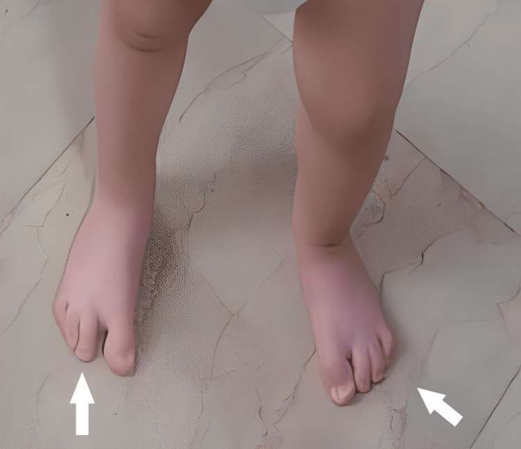
Figure 1. 1st patient’s feet at the age of 2 years.
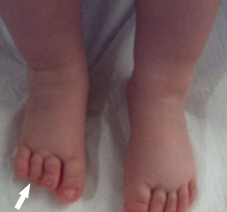
Figure 6. 2nd patient’s unilateral oligodactyly, with a marginally smaller foot, difficult to be spotted unless it was notified at the age of 1 year.
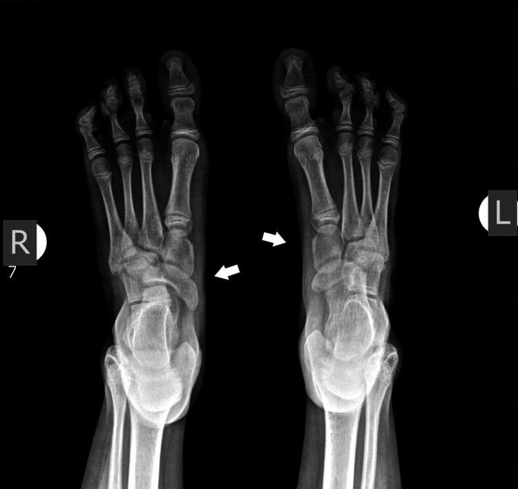
Figure 5. X-ray of 1st patient’s feet at the age of 7 years with an enlarged navicular, with a coalition of the talus navicular, and normal bilateral calcaneocuboid joint. There are two cuneiforms present.
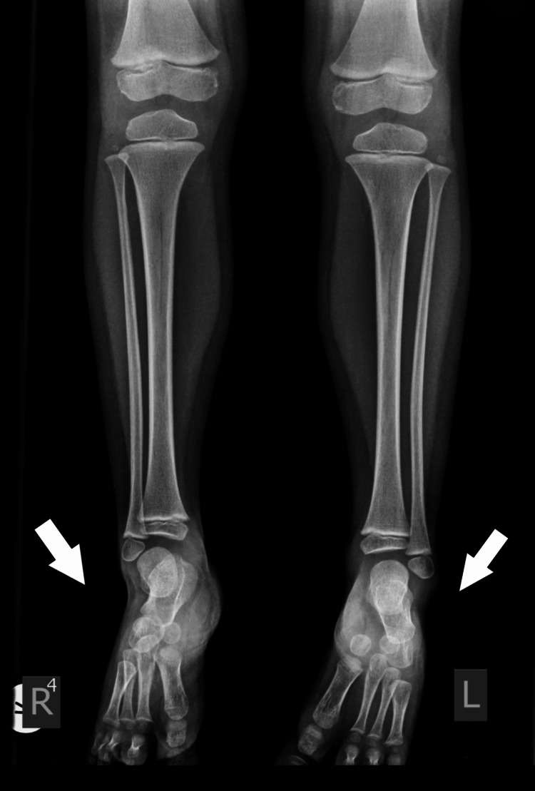
Figure 4. X-ray of 1st patient’s feet with bilateral involvement at the age of 2 years. Normal development of the tibia and fibula. Delayed initial ossification of the navicular.
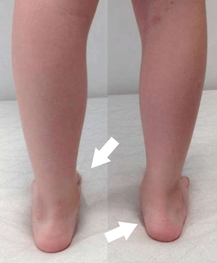
Figure 3. 1st patient’s feet at the age of 7 years – posterior view
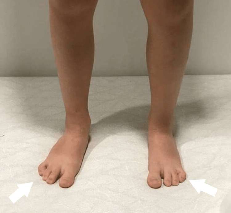
Figure 2. 1st patient’s feet at the age of 7 years – anterior view
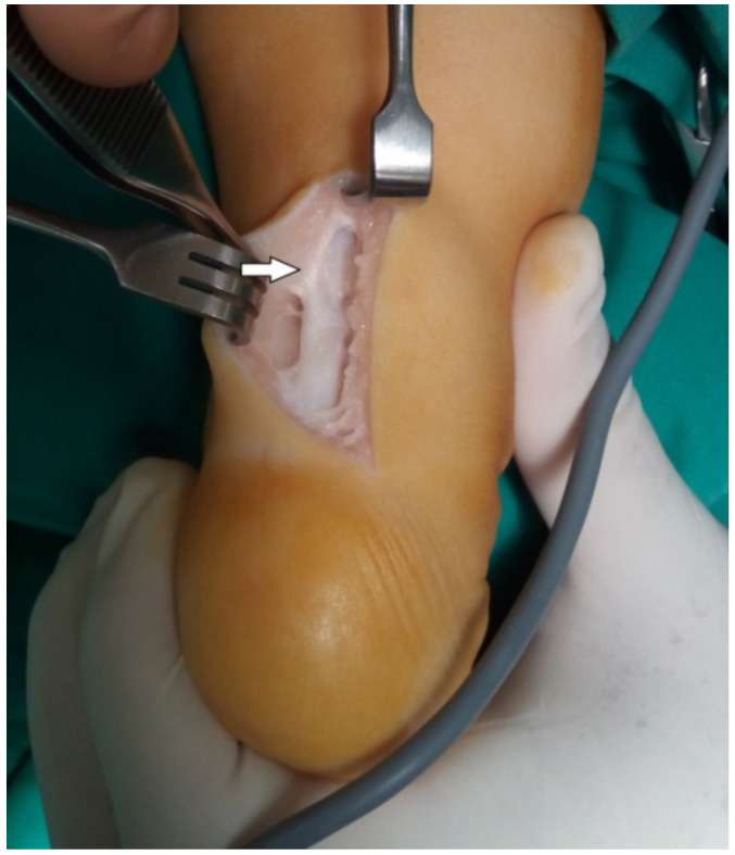 Anatomical Structures Responsible for CTEV Relapse after Ponseti Treatment