Myositis ossificans (MO) is a rare benign tumor characterized by heterotopic ossification of soft tissues. While the term “myositis” suggests that the lesion commonly affects muscles, it can also occur in rare locations such as tendons or subcutaneous tissues. MO is infrequent in children, primarily affecting young adults and adolescents. It presents distinct radiological and histological features. However, it is crucial to exclude malignancy when investigating heterotopic ossification. Surgical excision of the lesion following an appropriate biopsy is an option, but cases of self-limiting MO have been reported. We report a unique case of a 14-year-old girl who presented with painful restriction of movement in the lumbar and gluteal regions, without any history of trauma. The girl underwent thorough clinical and radiological investigations, with MRI revealing a circumscribed ossifying lesion measuring 3.8 × 2.3 cm in the gluteal subcutaneous area, lacking clear boundaries, with edema of the adjacent muscles. The lesion exhibited heterogeneous signal intensity and contrast enhancement, while 18F-FDG PET/CT revealed increased metabolic activity within the lesion. A US-guided needle biopsy was performed, confirming the diagnosis of MO with a typical zonal phenomenon. The girl was referred for surgical removal of the lesion within three months after the initial MRI. Upon re-evaluation, the mass was barely palpable, and subsequent MRI and CT scans showed a marked reduction in lesion size. We followed the patient, and in another three months, the mass had almost completely regressed. We aimed to report a unique case of an adolescent with an established diagnosis of MO, following appropriate radiological and histological evaluation. The rapid regression of the lesion suggests that MO can be managed conservatively in selected cases rather than through immediate surgery.
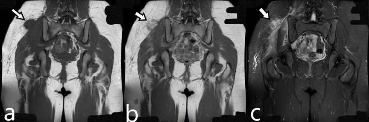
Figure 1. Coronal MRI images (a) Coronal T1W MRI shows a mass in the subcutaneous tissue with signal intensity similar to that of skeletal muscle, without clear boundaries and in contact with the gluteus muscle, measuring 3.8 × 2.3 cm. (b) Coronal contrast-enhanced T1W MRI shows an intensely heterogeneous enhancement of the mass. (c) Coronal FSE STIR MRI shows marked edema of the gluteus medius and minimus muscles and less intense edema in the paravertebral muscles that had the normal striation. FSE, fast spin echo
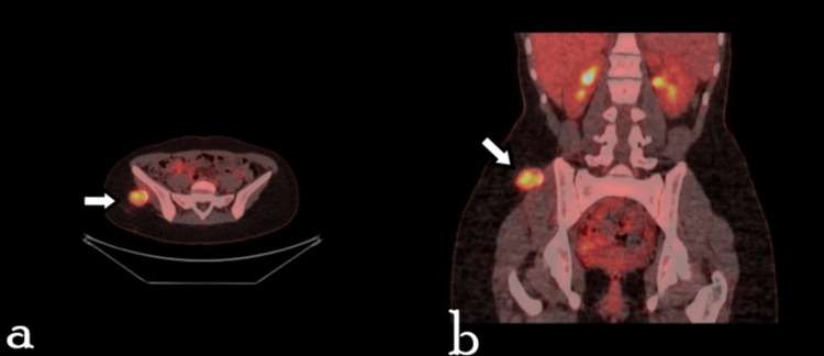
Figure 2. Transverse and coronal 18F-FDG PET/CT images show a hypermetabolic lesion within the adipose tissue, in contact with the gluteal muscle (a) Maximum intensity projection image shows a solitary hypermetabolic lesion within the soft tissues of the right gluteal region, along with axial CT (in bone and tissue windows), PET, and PET/CT sections. (b) The same sections shown in coronal view. The SUVmax of the lesion was 6.6, and the radiodensity measured 42 and 225 HU in the interior and at the calcified rim of the lesion, respectively (for comparison, the radiodensity of the cortical iliac bone was 560 HU).
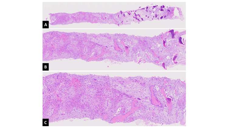
Figure 2. Transverse and coronal 18F-FDG PET/CT images show a hypermetabolic lesion within the adipose tissue, in contact with the gluteal muscle (a) Maximum intensity projection image shows a solitary hypermetabolic lesion within the soft tissues of the right gluteal region, along with axial CT (in bone and tissue windows), PET, and PET/CT sections. (b) The same sections shown in coronal view. The SUVmax of the lesion was 6.6, and the radiodensity measured 42 and 225 HU in the interior and at the calcified rim of the lesion, respectively (for comparison, the radiodensity of the cortical iliac bone was 560 HU).
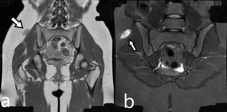
Figure 4. MRI images after two months (a) Coronal contrast-enhanced T1W MRI after two months from the initial MRI shows shrinking of the lesion, measuring 2.1 × 1 cm. (b) Coronal FSE STIR MRI obtained two months after the initial MRI shows reduced edema in the gluteal and paravertebral muscles. FSE, fast spin echo
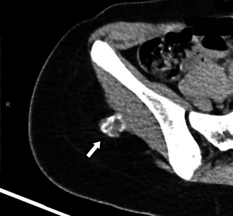
Figure 5. Axial CT scan after two months from the initial MRI shows a mass with peripheral ossifications
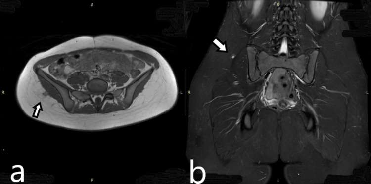
Figure 6. Axial contrast-enhanced T1W MRI and coronal FSE STIR images, after six months from the initial MRI, show a small lesion measuring 0.9 × 0.8 cm, with normal appearance of the surrounding muscles (a) Axial contrast-enhanced T1W MRI. (b) Coronal FSE STIR. FSE, fast spin echo