Introduction: Conservative management of femoral fractures in children in the preschool age is considered as the standard care. In the past years, there is an increase in the number of surgically treated children with IM nails, even in these young ages. Several papers report on the results comparing the final alignment of the fractures but there are no reports on the conservative treatment of a fracture with transposition of the fragments.
Case report: A 3-year-old boy was referred in our hospital with a spiral fracture of his right femur. There was transposition of the fragments, with the proximal part shifted medially and the distal part laterally. We informed the parents for the methods of treatment available for his fracture. After 1 week of hospitalization, the child was transferred home, with skin traction, with supervision of an orthopedic surgeon. The child was regularly evaluated with radiological examination, every 10 days. There was an uneventful healing of the fracture, with normal alignment of the leg and acceptable initial leg length discrepancy (LLD). One year after the fracture, there is an impressive remodeling of the fracture with restoration of the medullary canal and absent LLD.
Conclusion: Conservative treatment of spiral fracture in a child younger than 3 years is an effective method. Remodeling in this young age can restore not only deviations on the frontal and sagittal plane but also correct the transposition of the fragments with restoration of the medullary canal.
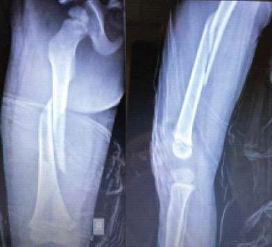
Figure 1 (a and b) Initial fracture, with transposition of the AP and good lateral alignment.
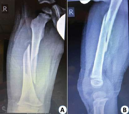
Figure 2 (a and b) X-ray examination, at 3 weeks, with ossification of the ends of the fractured parts of the femur.
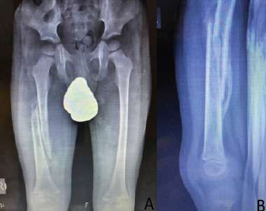
Figure 3 (a and b) X-ray evaluation at 4 weeks after injury with further increase of the healing process.
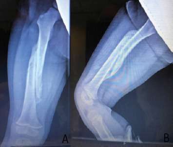
Figure 4 (a and b) Adequate bone formation of the fractured femur at 4 months.
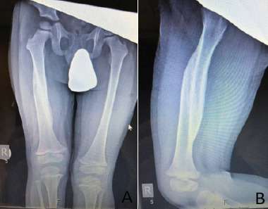
Figure 5 (a and b) At 1 year, there is an impressive correction of the shape of the femur, with restoration of the medullary canal, with the thin cortices line between the fragments.