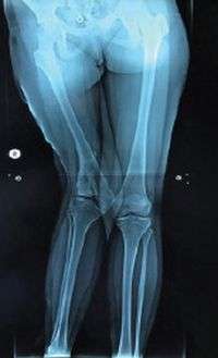
Fetuses with prenatal diagnosis of reduced mobility and malformation are at increased risk to sustain neonatal femoral fractures. Labor difficulties even after an elective cesarean section may trigger the fractures of the long bones.
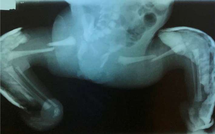
Figure 2 X‐ray showing bilateral femoral fractures. Casts were applied for fixed equinovarus feet
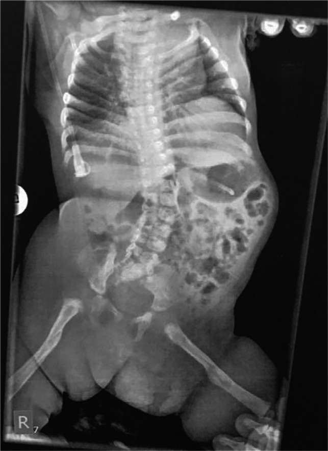
Figure 3 X‐ray showing complete healing of femoral fractures, improvement of scoliosis, and pelvic obliquity
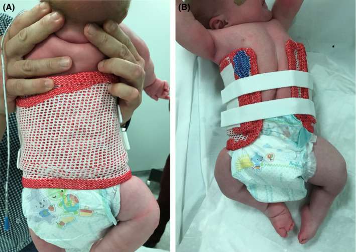
Figure 4 A and B, Cast for the correction of the spine scoliosis and pelvic obliquity posterior and anterior view
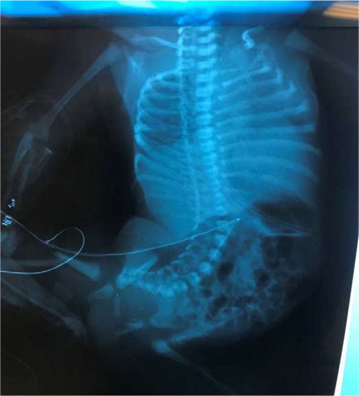
Figure 1 X‐rays soon after birth showing severe fixed scoliosis, pelvic obliquity, and bilateral femoral fractures