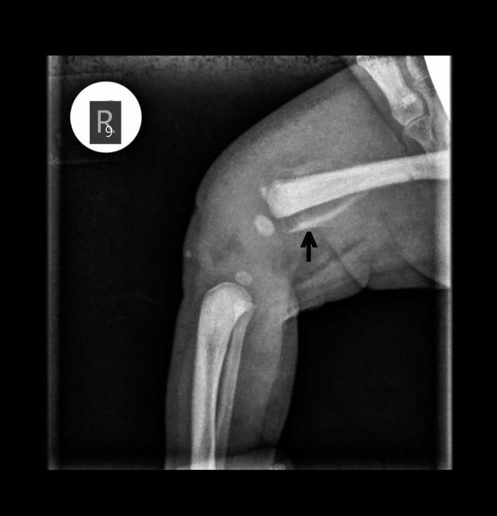
Localized pain in the absence of local lesion may represent referred pain from the spine, in a child, arising from a benign spinal schwannoma. It can be diagnosed by MRI. Surgical excision of the tumor relieves the symptoms.
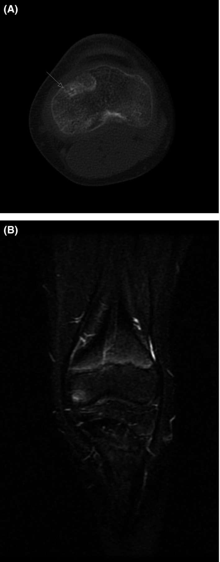
Figure 1 A‐B: Initial MRI and CT scan with an area of lucency with edema that led to incorrect diagnosis for a possible OO
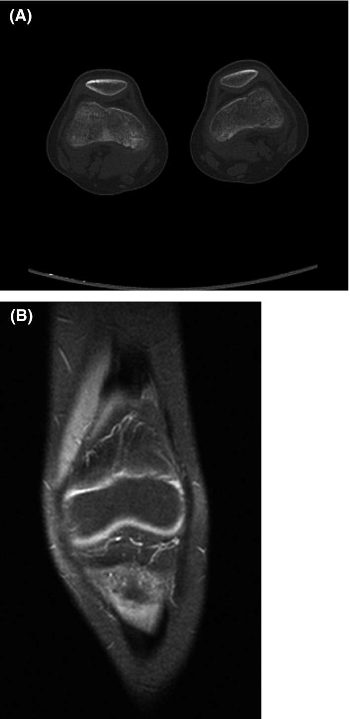
Figure 2 A‐B: Our CT and MRI examination with normal findings
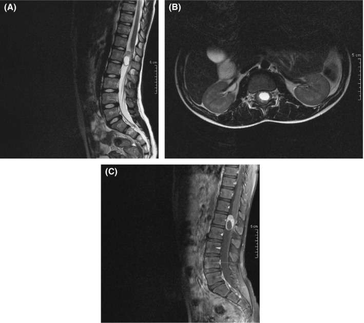
Figure 3 A, Sagittal T2W sequence. An oval intradural extramedullary lesion is observed at the level of the L1 vertebra. The central cystic part of the lesion demonstrates high signal in T2W sequences, whereas the peripheral solid part appears with intermediate signal. B, Axial T2W sequence. In the transverse plane, the lesion appears to be located in the central and left part of the thecal sac, causing pressure to the cauda equina. C, Sagittal T1W + C. After the intravenous administration of paramagnetic agent, there is vivid enhancement of the peripheral solid part of the tumor, with the central cystic component of the lesion remaining unenhanced
show full caption
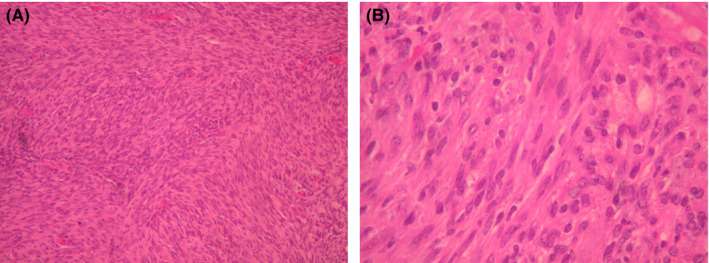
Figure 4 A, The tumor shows increased cellularity and the tumor cells are spindle‐shaped and arranged in fascicles. B, Tumor cells without nuclear atypia or mitosis