A rare clinical presence of Salter-Harris type II fracture of the distal femur in a newborn. The crucial role of imaging in depicting urgent anatomical alterations.
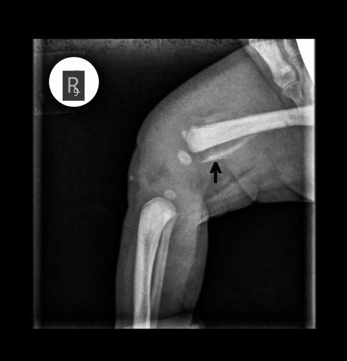
Figure 1 X‐ray of the right femur. The black arrow indicates the fracture and the detached osseous part
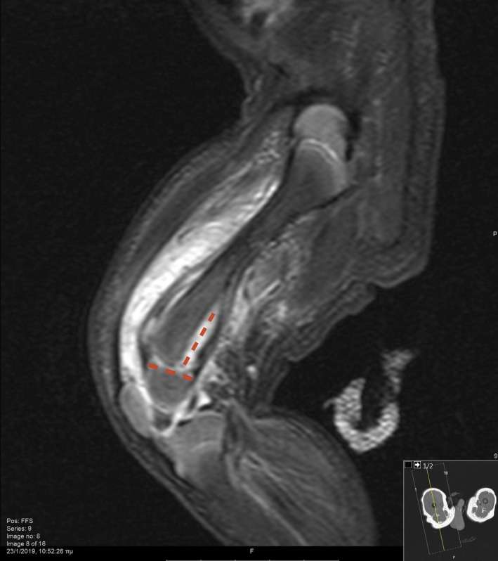
Figure 2 MRI of the right femur depicting the presence of hematoma and disruption of periosteum, setting the diagnosis of Salter‐Harris type II fracture. The red lines indicate the type of the fracture