A calcified mass located in the subcutaneous area above the Achilles tendon of a child poses difficulty for a correct diagnosis. Differential diagnosis includes tumoral calcinosis, calcified fibroma, calcified tendinitis, and myositis ossificans. A fibro-osseous pseudotumor is a rare benign lesion that usually affects the digits. Histologically, it consists of fibrous tissue with areas of osteoid formation. We report a child with a hard subcutaneous mass above the Achilles tendon. Radiographic assessment, including X-ray, MRI, and CT scan, revealed a calcified tumor in the posterior part of the Achilles tendon, in proximity to the sural nerve and vessel. After surgical removal of the mass, histopathology revealed fibrous connective tissue with areas of islands and trabeculae of osteoid, confirming the diagnosis of a fibro-osseous pseudotumor. A subcutaneous calcified mass in a child requires appropriate clinical and pathological investigation to ensure an accurate diagnosis.
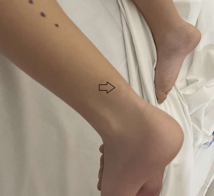
Figure 1. Clinical picture with the arrow pointing to the subcutaneous swelling.
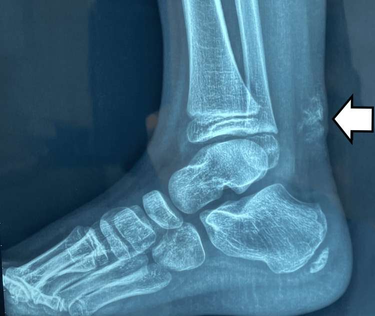
Figure 2. X-ray showing irregular calcification in the subcutaneous tissue, overlying the Achilles tendon.
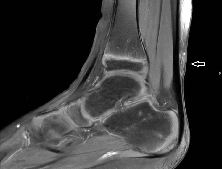
Figure 3. MRI scan on the sagittal plane showing a soft tissue subcutaneous mass overlying the lower third of the Achilles tendon, with areas of lower signal intensity on T1. The image demonstrates calcified tissue with mild enhancement after contrast medium administration
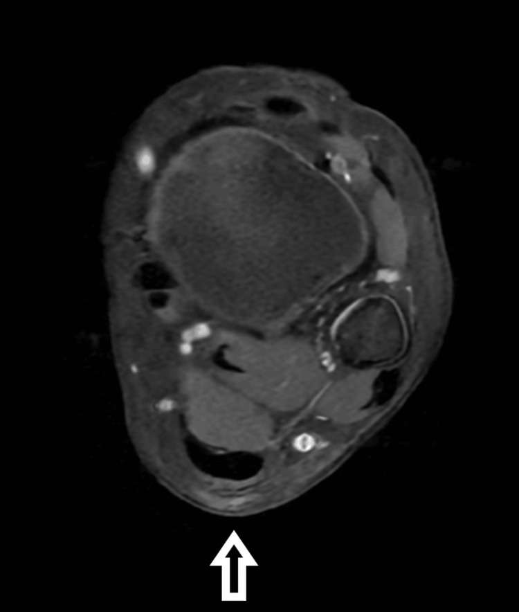
Figure 4. MRI scan on the transverse plane showing a soft tissue subcutaneous mass overlying the lower third of the Achilles tendon, with areas of lower signal intensity on T1.
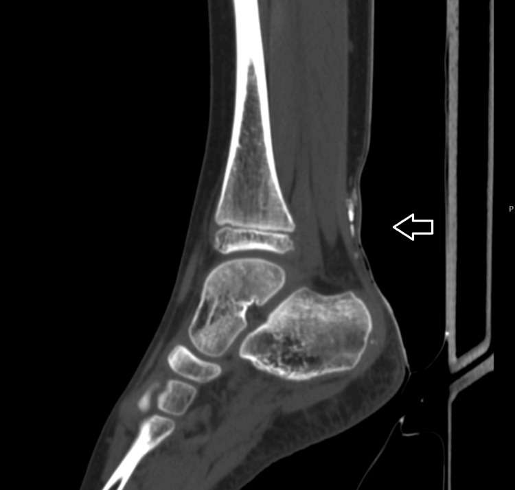
Figure 5. CT image on the sagittal plane showing a thin calcified lesion, located above the Achilles tendon, extending toward the sural nerve and vessel.
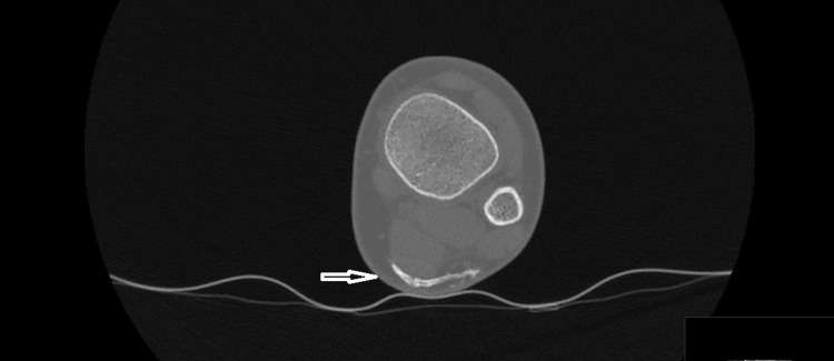
Figure 6. CT image on the axial plane showed a thin calcified lesion, located above the Achilles tendon, extending toward the sural nerve and vessel.