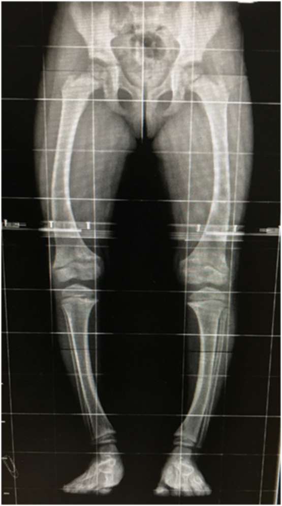
An aneurysmal bone cyst (ABC), when located juxta-physeal, may rarely penetrate the growth plate and extend into the epiphysis. The recurrence rate is considered higher when ABC is in contact with the active growth plate. Treatment methods usually focus on cyst healing and the rate of cyst recurrence. We present the method of treatment used for addressing the lesion of the growth plate following the surgical management and healing of a juxta-physeal ABC. A seven-year-old girl had an aggressive ABC in the juxta-physeal area of the distal femur, penetrating the growth plate and extending in the epiphysis. Surgical treatment was performed, including curettage and autologous bone grafting, avoiding the growth plate. The cyst healed; however, physis presented an obliteration. The affected limb developed valgus deformity and severe leg length discrepancy (LLD). To address this issue, once our patient completed her growth, we proceeded with distraction osteogenesis, using the Ilizarov device, with asymmetrical lengthening of the rods. We achieved the correction of the limb alignment and resolved the LLD. ABCs in the juxta-physeal area of a growing child are benign metaphyseal tumors that exceptionally may penetrate the physis and extend into the epiphysis. Our report highlights that the growth plate’s lesion, despite the cyst’s healing, may compromise the final result. The use of the Ilizarov device is an effective method for correcting the malalignment and the LLD that may emerge.
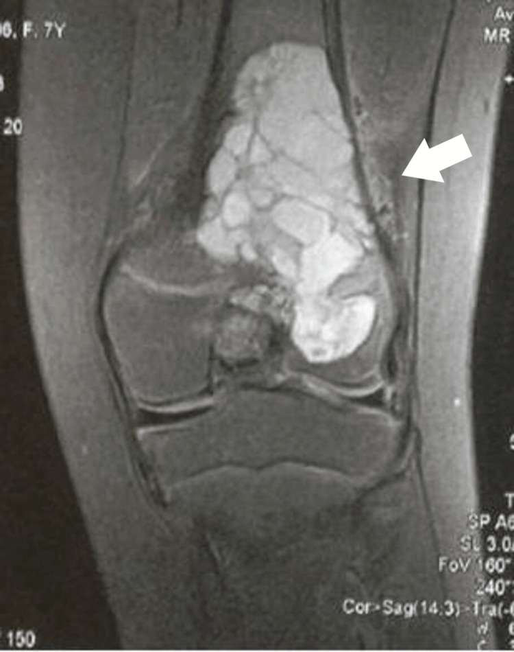
Figure 1. A coronal T1-weighted MRI image showing multiple fluid-fluid levels in the metaphysis, crossing the physis, and extending into the epiphysis.
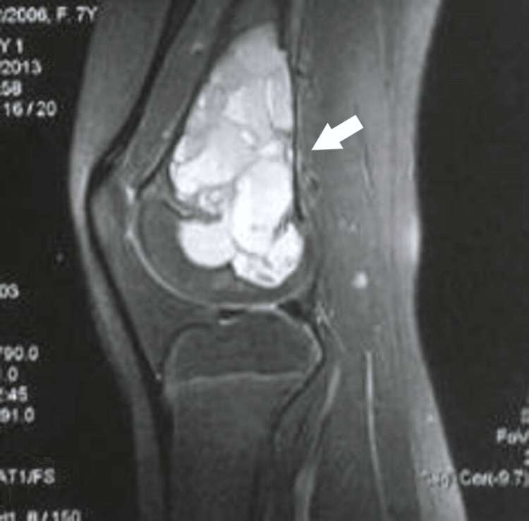
Figure 2. A sagittal T1 FS (Gadolinium-enhanced) weighted MRI image displaying multiple fluid-fluid levels in the metaphysis, traversing the physis and progressing into the epiphysis. Observe the thinning of the posterior cortex and the lack of a periosteal reaction. FS: Fat saturation.
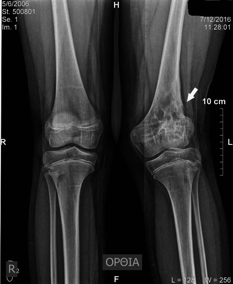
Figure 3. Standing X-ray image illustrates the healing process of the cyst and depicts the restored cortex. A reduced height of the lateral femoral condyle and obliteration of the normal radiolucency of the physis are also observable.
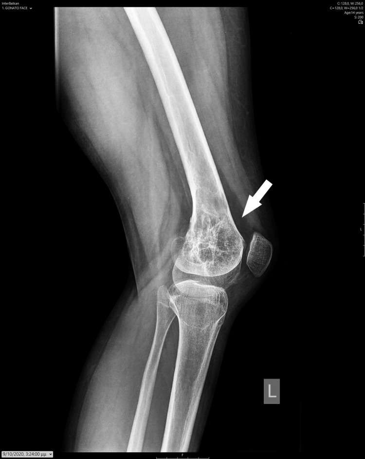
Figure 4. Lateral X-ray image depicts the healed cyst and the restored cortex.
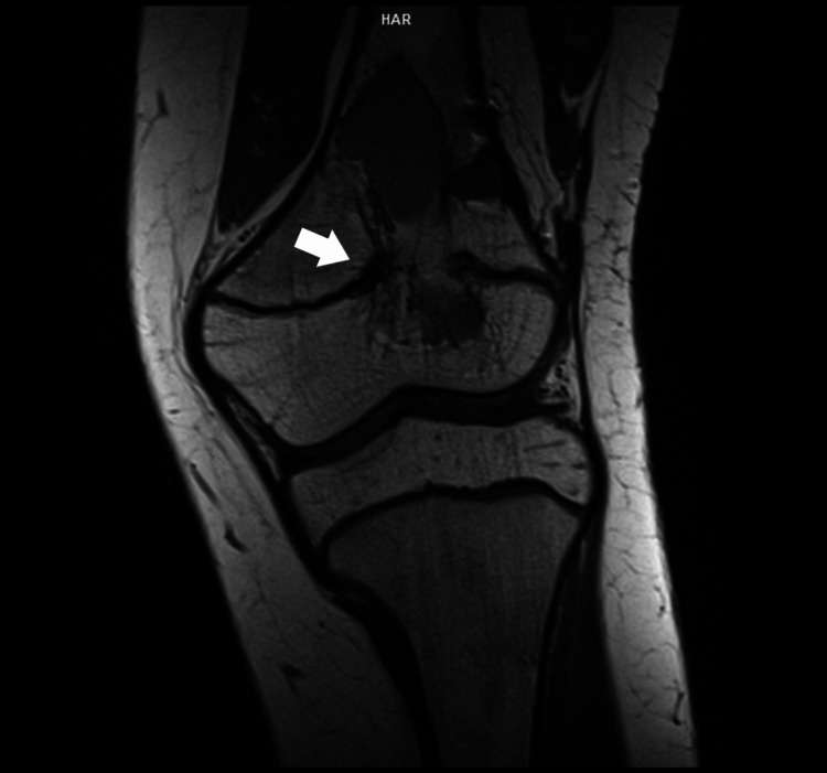
Figure 5. Coronal T1 MRI image displays the diminished diameter of the remaining cyst elements, alongside disturbance of the normal growth plate line.
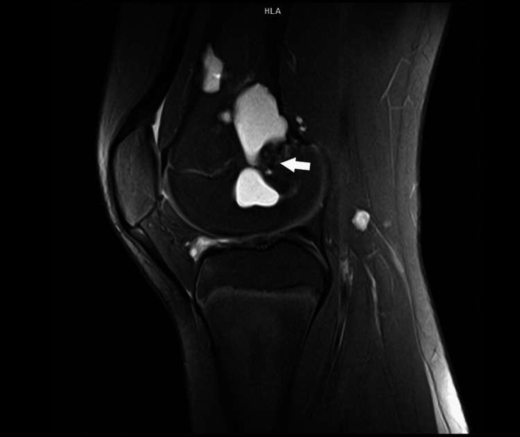
Figure 6. Sagittal T1 FS MRI image displays the reduced diameter of the remaining elements of the cyst, along with the disturbance of the normal line of the growth plate. FS: Fat saturation.