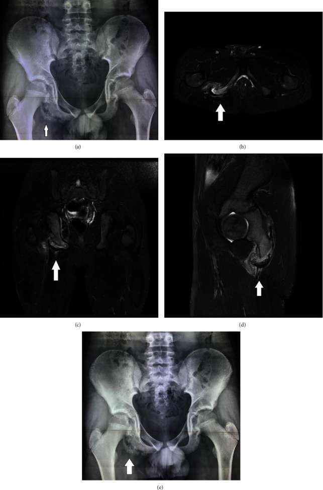
Introduction: Solitary osteochondromas are extremely rare in the bones of the foot. In the growing skeleton, few cases affecting the metatarsals and the talus have been reported. At present, there have been no reports of osteochondromas affecting the cuneiforms.
Case report: We report the case of a 13-year-old male patient. He presented with marked prominences in the plantar surface of his left foot and pain while participating in sporting activities. Radiological examination with X-rays, computed tomography (CT) scan, and magnetic resonance imaging revealed two solitary osteochondromas growing from the medial cuneiform and the head of the 1st metatarsal. The patient was treated surgically by excision of the osteochondromas. Histological examination confirmed the diagnosis of osteochondromas. He had an uneventful recovery and returned to his sporting activities.
Conclusion: Solitary osteochondroma can present in the cuneiform and metatarsal of a growing adolescent. CT scan is useful for the accurate diagnosis and surgical removal of the tumor.
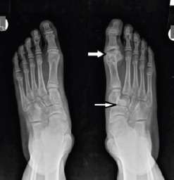
Figure 1 X-ray of feet: On the right foot, there is an osteochondroma in the lateral area off the 1st metatarsal, with enlargement of the MTF joint. Increased ossification is noticed in the area of the middle cuneiform.
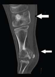
Figure 2 CT scan sagittal: Osteochondromas of the cuneiform and metatarsal. The osteochondroma is communicating with the medullary canal of the metatarsal and the cuneiform.
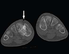
Figure 3 CT scan axial both feet: The osteochondroma arising from the medial cuneiform, with calcification, extending laterally and plantar.
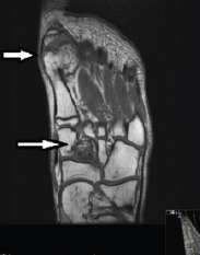
Figure 4 Coronal T1-weighted MRI reveal the cortex-medulla continuation between osteochondromas and parental bones (1st metatarsal and medial cuneiform). The cortex displayed low signal intensity and the medulla high signal intensity on T1W images.
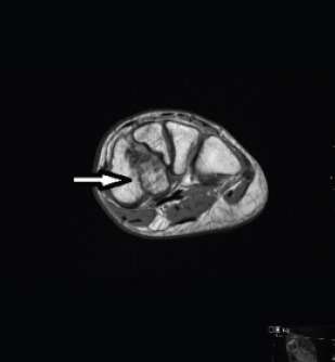
Figure 5 STIR axial, at the level of the 1st metatarsal. The thin hyperintense hyaline cartilage is easily assessed in STIR images, while on T1W images, it appears with intermediate signal intensity.
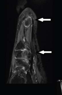
Figure 6 Axial T1W image at the level of cuneiforms: Typical imaging findings of osteochondroma of the medial cuneiform with thin hyaline cartilage cap.