Solitary extraskeletal osteochondromas are rare benign lesions usually located close to a joint and are characterized by the absence of continuity with the adjacent bone. They are usually found in the hand and feet and are extremely rarely reported in the growing skeleton. In this paper, we describe a four-year-old boy who presented with a solitary calcified tumor in the posterior part of his ankle. We performed a detailed evaluation using plain X-rays, a CT scan, and an MRI, which revealed a well-demarcated calcified tumor that had the characteristics of an osteochondroma but without any continuity with the bones of the ankle joint. The lesion was treated surgically with the excision of a giant osteochondral lesion. Pathological examination revealed mature cartilage at the periphery with cancellous bone in the central part. Thus, we present the clinical and laboratory investigation of a solitary extraskeletal osteochondroma in the ankle of a four-year-old boy, which is an extremely rare case.
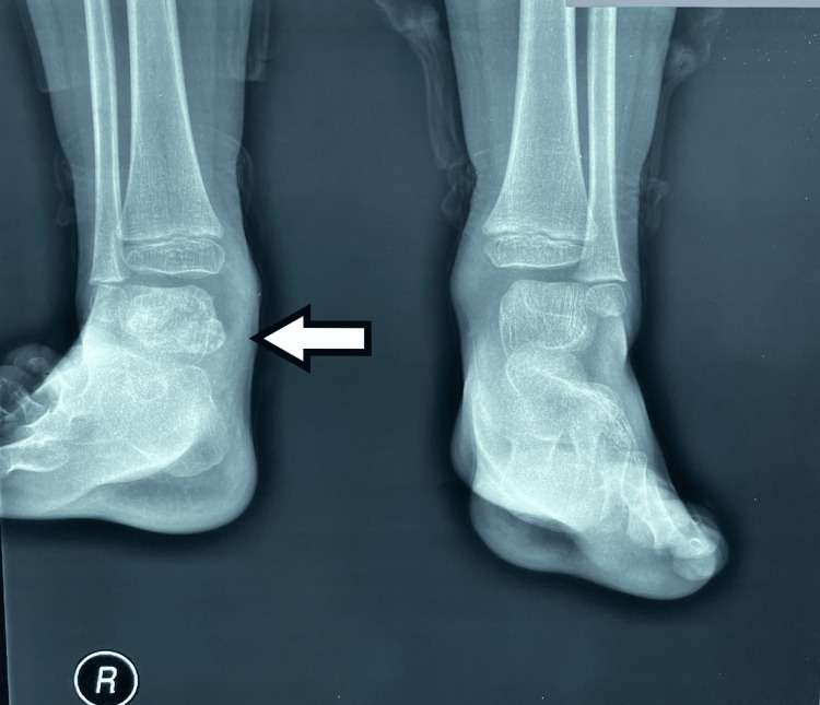
Figure 1. Plain radiographs of the ankle joints in anteroposterior projection. A large calcified lesion, with lobulated margins, is observed adjacent to the posterior margin of the right talus.
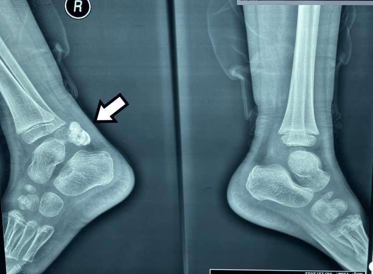
Figure 2. Plain radiographs of the ankle joints in lateral projection. A large calcified lesion, with lobulated margins, is observed adjacent to the posterior margin of the right talus.
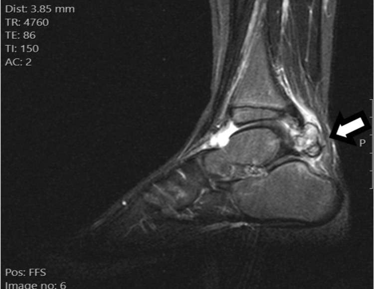
Figure 3. MRI scan of the right ankle sagittal STIR sequence. Short tau inversion recovery (STIR) sequence in the sagittal plane demonstrates a large lobulated mass in the posterior aspect of the ankle joint, which projects in the Kager’s fat pad. The mass has an inhomogeneous appearance, with interchanging areas of high and low magnetic resonance signals. The lesion is in close proximity to the cortex of the posterior surface of the talus, without clear evidence of a bony protuberance that connects the lesion with the host bone. An increased amount of fluid is observed in the ankle joint.
show full caption
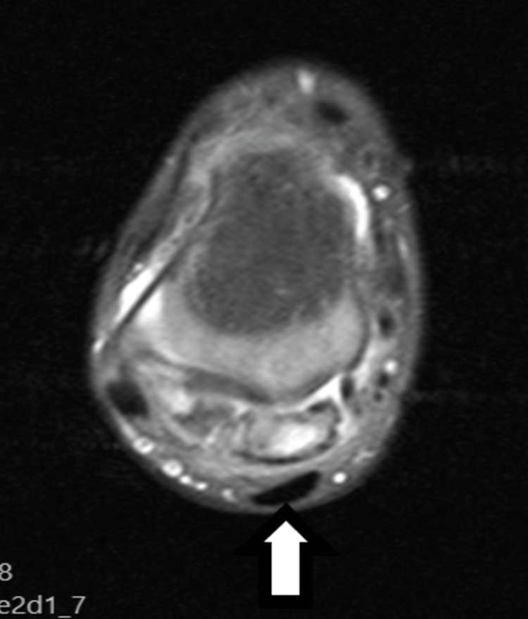
Figure 4. MRI scan of the right ankle, axial proton density (PD) fat saturated. Proton density (PD) sequence with fat suppression in the axial plane also demonstrates the relationship of the mass with the adjacent anatomic structures.
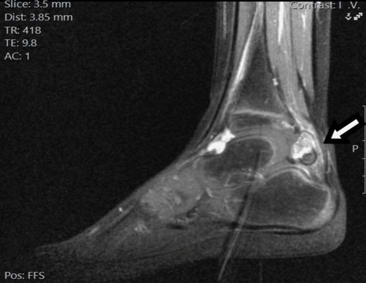
Figure 5. MRI scan of the right ankle T1C sequence. T1C sequence in the sagittal plane after the intravenous administration of paramagnetic agent shows inhomogeneous enhancement of the lesion.
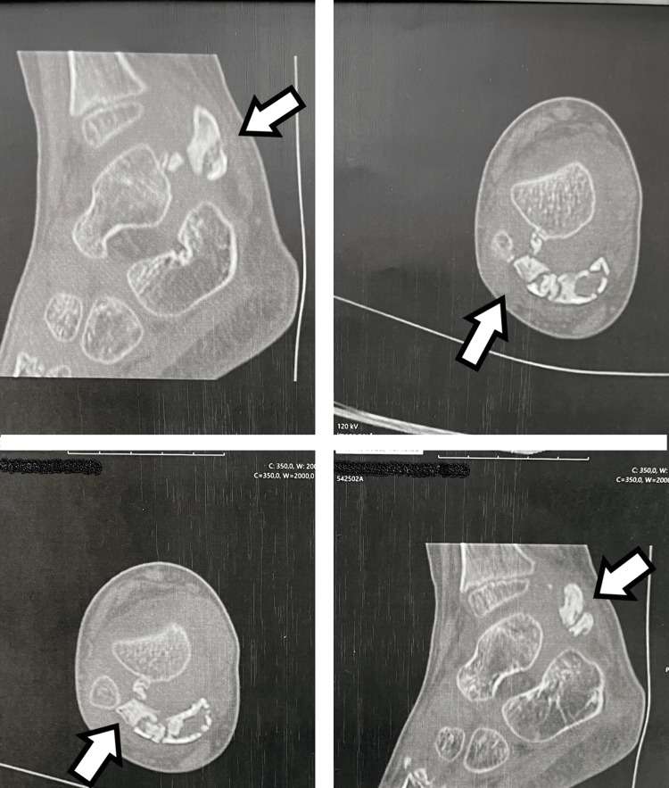
Figure 6. CT scan of the right talus with multiplanar reconstruction. There is extensive calcification of the mass. A small calcified stalk is observed between the lesion and the talus, but no clear continuity of the stalk with the talus is confirmed.