Osteochondritis of the ischiopubic area is a rare disease of children that presents with hip pain and limping. Careful examination and appropriate investigations are essential to establish a definite diagnosis. We report a case series of four children, ages 10-14-year-old, with osteochondritis of the ischiopubic area. Plain X-ray examination showed an area of diffuse irregular calcification of the ischium in two of the children, while in the other two there was an asymmetrical enlargement of the ischiopubic synchondrosis. MRI investigation was the most helpful examination. Bone edema was found in all four children. A calcified mass separated from the host ischium was found in the first two children. The cortex was normal, without irregular destruction. Bone edema of both the ischium and pubic alongside the synchondrosis was found in the following two children, with intact cortices and asymmetrical enlargement. Osteochondritis lesions of the ischium and the ischiopubic area have radiological findings similar to several severe diseases. Bone edema on MRI investigation in children must be properly evaluated. Appropriate radiological examination enabled us to confirm the diagnosis of the osteochondritis and to avoid unnecessary procedures. We want to draw attention to the rare diagnosis of osteochondritis of the ischiopubic area, and the clinical significance, as a cause of hip pain and limping in children.
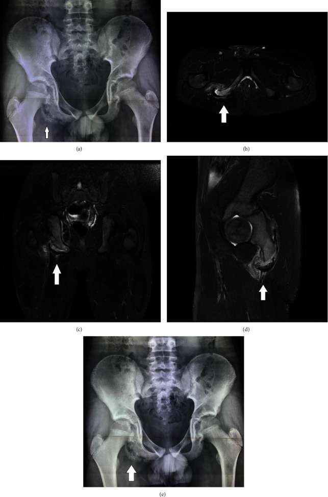
Figure 1 (a) On the right ischium, there is calcification adjacent to the parental bone that has normal structure. (b–d) STIR sequence in transverse (b), coronal (c), and sagittal (d) plane of the pelvis in an adolescent with right hip pain. An asymmetric enlargement of the right ischial tuberosity is observed, with a semilunar osteocartilaginous mass separated from the body of the ischial tuberosity. A fluid field cavity is developed between the parental bone and the detached fragment, whereas concomitant edema of the surrounding muscles due to blood products is also observed. The conjoined hamstring tendon remains attached to the apophysis. (e) There is partial union of the calcified osteochondral bone to the ischium.
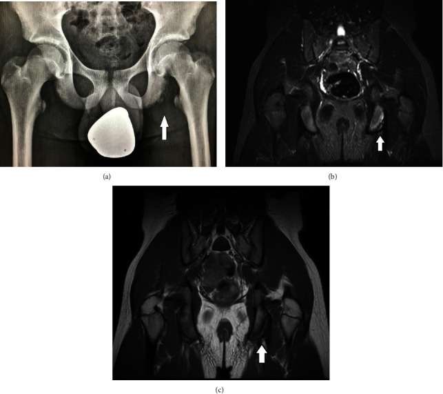
Figure 2 (a) There is an irregular cortex of the left ischial tuberosity, with a smooth line of calcification, in close proximity to the host bone. STIR (b) and T1W (c) sequences in coronal plane of an adolescent with left gluteal pain demonstrate an avulsion lesion of the left ischial tuberosity, with edema of the ischium and minimal displacement of the osteochondral lesion.
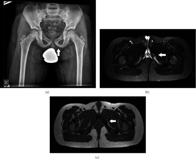
Figure 3 (a) Asymmetric enlargement of the left ischiopubic synchondrosis. (b and c) STIR (b) and T1W (c) sequences in transverse plane in an adolescent with left gluteal pain, demonstrate irregularity of the left ischiopubic synchondrosis, with bone marrow edema involving the bony structures around the synchondrosis. The adjacent soft tissues maintain their normal MRI signal.
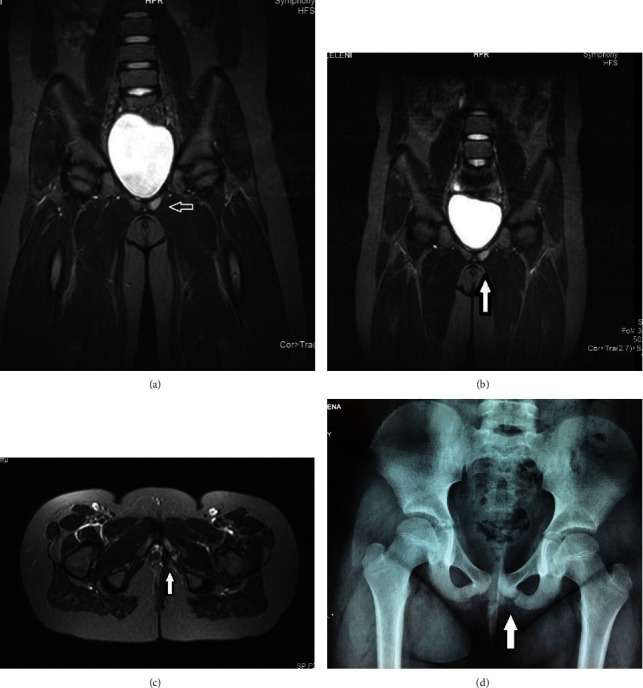
Figure 4 (a–c) STIR sequences in (a and b) coronal and (c) transverse plane demonstrate bone marrow edema of the left ischium, with normal appearance of the cortex and normal appearance of the adjacent muscles. (d) Asymmetric enlargement and diastasis of the left ischiopubic synchondrosis.