Perivascular epithelioid cell tumors (PEComas) are rare mesenchymal tumors composed of cells with epithelioid and spindle morphology, which grow around blood vessels. PEComas of soft tissue are usually reported in adults, with a rare incidence in the pediatric population, especially in young children and in the soft tissue of the limb. We report a case involving a healthy seven-year-old girl who presented with a painless prominence in front of the tibial tubercle. Radiological imaging revealed an encapsulated, lobulated mass that simulated a lipoma. After surgical excision, the microscopic examination and histopathological analysis of the surgical specimen confirmed the diagnosis of benign PEComa. Our case underscores the importance of considering PEComa in the differential diagnosis of pediatric soft tissue masses in children, whose presentation may vary from benign to malignant. Surgical treatment is the preferred approach for benign PEComas.
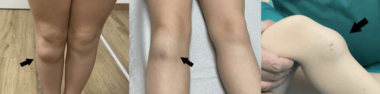
Figure 1. Prominence in the area of the tibial tubercle presenting as a painless mass (arrows).
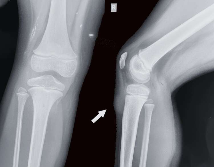
Figure 2. X-ray of the knee demonstrating a clear margin soft tissue enlargement, with intact bone and no periosteal reaction (arrow).
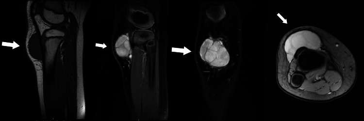
Figure 3. MRI images show an oval lesion on subcutaneous area in front of the tibial tubercle with increased signal intensity on T2-weighted images and low signal intensity on T1-weighted images. There are thin internal septa with smooth external contour and no bone involvement. There is absence of surrounding edema (arrows).
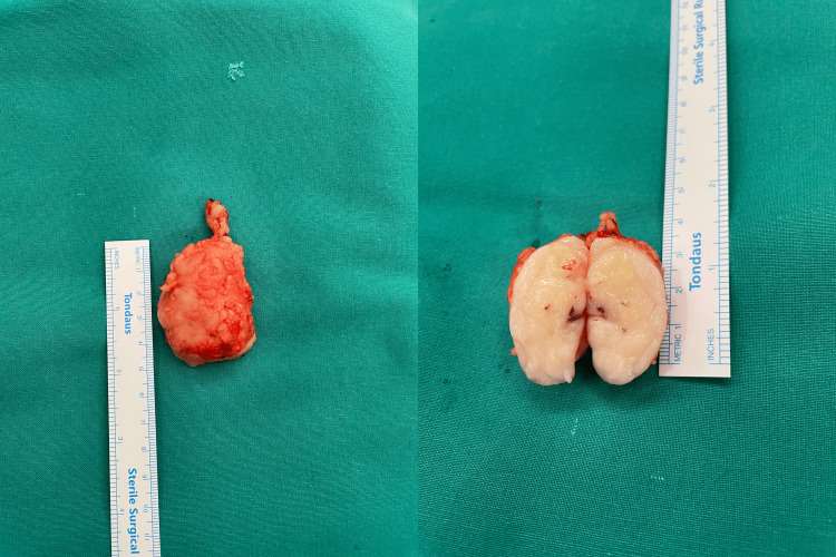
Figure 4. The mass is marginally 5 cm in diameter, appearing firm and elastic, giving the impression of a lipoma.
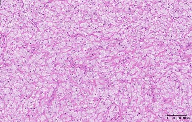
Figure 5. The tumor is composed of large epithelioid cells with compressed capillary vessels between them in a solid arrangement (hematoxylin and eosin (H&E) stain, ×10).
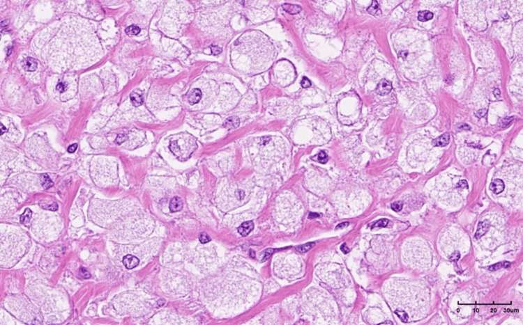
Figure 6. Epithelioid cells with clear, finely granular cytoplasm, with mild atypia and rare mitosis (hematoxylin and eosin (H&E) stain, ×40).