Introduction: Congenital distal tibiofibular diastasis is an extremely rare disorder. The main feature at presentation is a clubfoot deformity with leg length discrepancy (LLD) due to a short tibia. Limb lengthening and reconstruction techniques today can correct the deformity, ending with a plantigrade and functional limb.
Case report: We report a patient with congenital tibiofibular diastasis, who was initially treated with series of casts and later with ankle arthrodesis, but the deformity and LLDleg length discrepancy remained. We applied an Ilizarov frame and initially corrected the varus and equinus deformity of the foot. Then, we corrected the LLD leg length discrepancy with limb lengthening with distraction osteogenesis. We achieved a plantigrade stable limb, with a small amount of length inequality.
Conclusion: Congenital distal tibiofibular diastasis can be managed with staged correction and lengthening with the Ilizarov method.
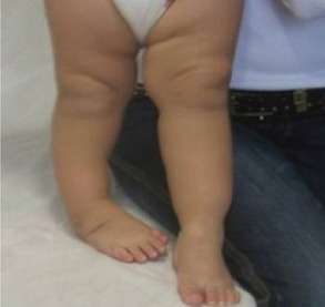
Figure 1 Initial presentation as clubfoot deformity. Clinical picture with clubfoot and shortening of the limb.
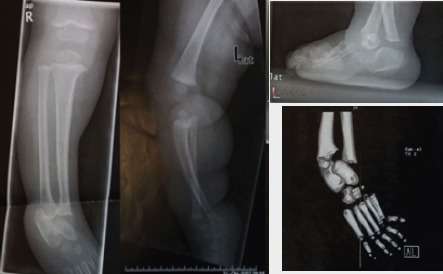
Figure 2 Distal tibiofibular diastasis, initial X-ray, and computerized tomography (CT) scan. Congenital distal tibiofibular diastasis, with the talus intervening, with a CT reconstruction.
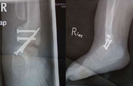
Figure 3 Arthrodesis of distal tibiofibular diastasis, X-ray. X-ray after the performed arthrodesis, with the attempt to correct the deformity.
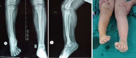
Figure 4 X-ray AP both tibia and lateral X-ray of the affected limb, clinical picture, before surgical reconstruction. Radiological examination before the surgical reconstruction with the Ilizarov frame. Equinus and varus position of the foot. Limb length discrepancy.
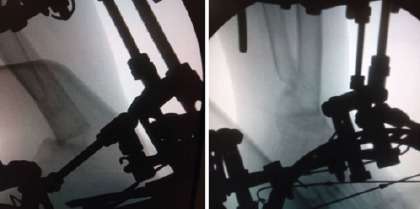
Figure 5 Correction of equinus and varus position with the Ilizarov frame. Opening wedge osteotomy and correction of equinus and varus with gradual distraction, with the Ilizarov frame
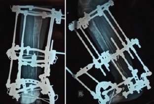
Figure 6 X-ray at the removal of the frame. Radiological examination with appropriate bone regenerates at the lengthening of the tibia.