
Osteoid osteoma (OO) most commonly affects the cortices of long bones and rarely involves the toes, where it typically presents with swelling and pain. Radiological evaluation can often pose a diagnostic challenge. We report the case of a boy with a painless swelling of the middle phalanx of the third toe. Radiographic assessment using plain X-rays, MRI, and CT revealed a centrally located sclerotic lesion with a barely visible nidus. A bone scan demonstrated increased uptake at the lesion site. Surgical intervention was performed to remove the sclerotic lesion, followed by reconstruction of the phalanx using an autologous bone graft. Histopathological analysis confirmed the diagnosis of OO. Although OO of the toes is uncommon, it may present as painless swelling. A centrally located sclerotic lesion in the phalanx warrants thorough investigation and surgical management with pathological confirmation to ensure accurate diagnosis.
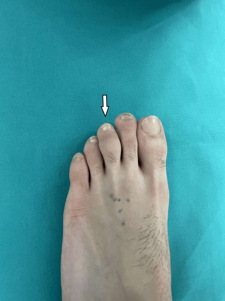
Figure 1. Swelling of the middle phalanx of the third toe, with normal skin coloration
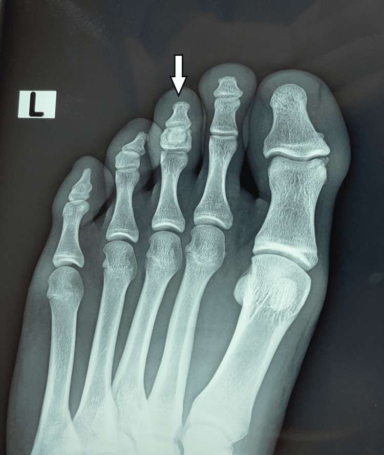
Figure 2. AP X-ray showing a central sclerotic lesion in the middle phalanx of the third toe, surrounded by a thin radiolucent zone
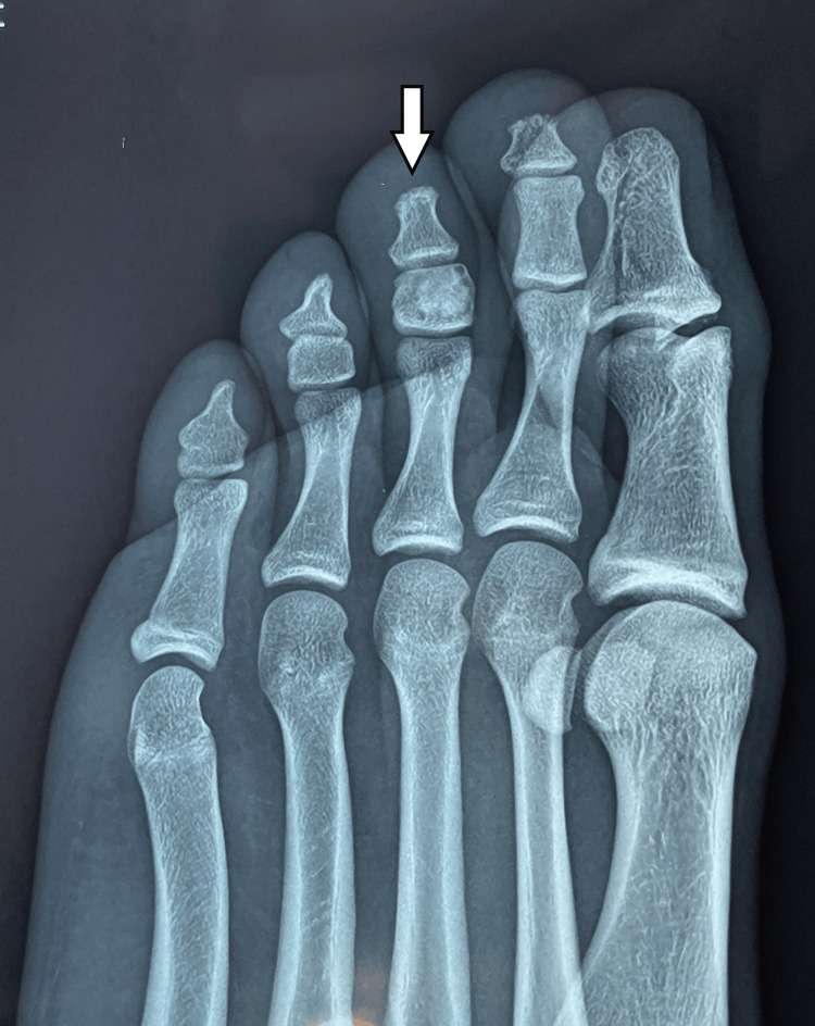
Figure 3. Lateral X-ray showing a central sclerotic lesion in the middle phalanx of the third toe, surrounded by a thin radiolucent zone
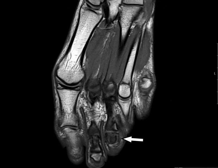
Figure 4. T1-weighted MRI showing an intermediate signal at the center of the lesion with low-signal intensity at the periphery
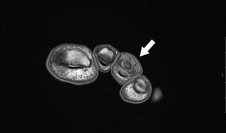
Figure 5. T1-weighted MRI showing an intermediate signal at the center of the lesion with low-signal intensity at the periphery
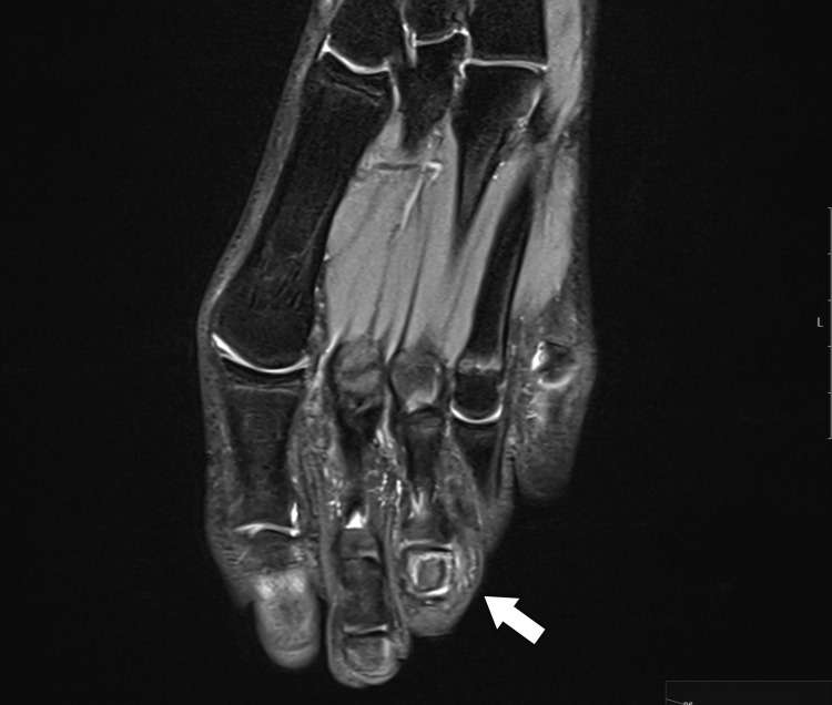
Figure 6. Axial T2-weighted MRI showing a centrally located high-intensity signal within the middle phalanx of the third toe, with a thin surrounding area of edema