orthopedikos

About admin

This author has not yet filled in any details.
So far admin has created 51 blog entries.

Melorheostosis in an Adolescent with Limb Length Discrepancy and Management with Epiphysiodesis with Eight Plates

2026-07-08T12:58:33+03:00

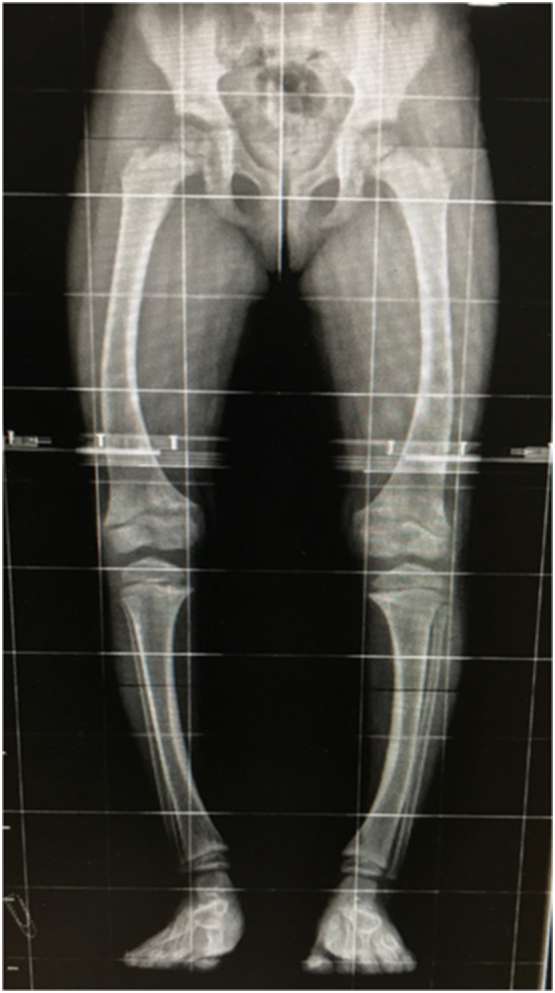
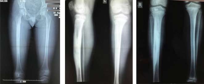
Introduction: Melorheostosis is a rare bone dysplasia. It is mainly a radiographic diagnosis with distinct findings of endosteal or periosteal sclerosis along the axis of long bones, resembling the flowing wax. It is usually polyostotic and monomelic. Clinical manifestations are pain, limitation of joint movements, deformities, and contractures. Leg length discrepancy may be found, usually [...]
Melorheostosis in an Adolescent with Limb Length Discrepancy and Management with Epiphysiodesis with Eight Plates2026-07-08T12:58:33+03:00

Impressive Remodeling of the Fractured Femur with Transposition of the Fractured Parts, Treated Conservatively with skin Traction, in a 3-Year-Old Boy

2026-07-08T09:55:06+03:00

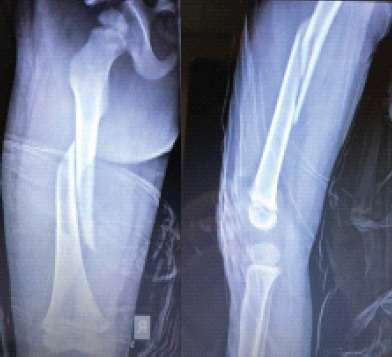
Introduction: Conservative management of femoral fractures in children in the preschool age is considered as the standard care. In the past years, there is an increase in the number of surgically treated children with IM nails, even in these young ages. Several papers report on the results comparing the final alignment of the fractures but [...]

Impressive Remodeling of the Fractured Femur with Transposition of the Fractured Parts, Treated Conservatively with skin Traction, in a 3-Year-Old Boy2026-07-08T09:55:06+03:00

Displaced Distal Fibular Epiphysis in a Child: A Clinical and Radiological Evaluation of a Rare Injury Hidden From the Initial Radiological Examination

2026-07-08T09:53:02+03:00

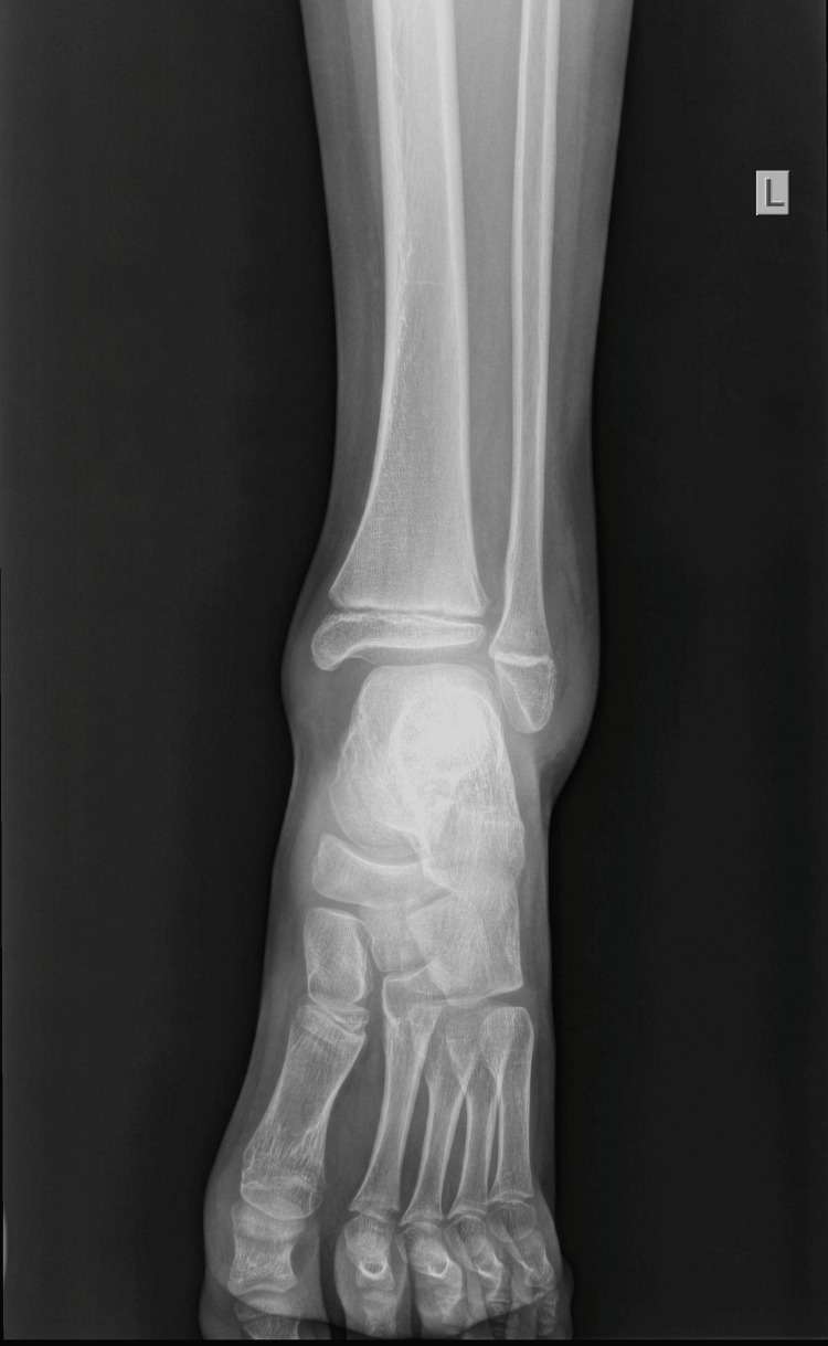
Fractures of the lateral malleolus are common in children. Isolated lesions of the distal fibula physis commonly comprise nondisplaced or minimally displaced fractures. An isolated, completely displaced epiphysiolisthesis of the distal fibula is an extremely rare lesion. This study introduces the case of an 11-year-old boy presenting an extremely rare lesion of an isolated, completely [...]

Displaced Distal Fibular Epiphysis in a Child: A Clinical and Radiological Evaluation of a Rare Injury Hidden From the Initial Radiological Examination2026-07-08T09:53:02+03:00

X-linked hypophosphatemia due to a de novo novel splice-site variant in a 7-year-old girl with scaphocephaly, Chiari syndrome type I and syringomyelia

2026-07-08T09:51:10+03:00

X-linked hypophosphatemia (XLH) is a rare X-linked dominant inherited disorder caused by loss-of-function variants in the PHEX gene and characterized by renal phosphate wasting, hypophosphatemia, abnormal vitamin D metabolism, growth retardation and lower limb deformities. We describe a case of XLH-rickets in a 7-year-old girl with scaphocephaly, Chiari syndrome type I and syringomyelia, with a [...]

X-linked hypophosphatemia due to a de novo novel splice-site variant in a 7-year-old girl with scaphocephaly, Chiari syndrome type I and syringomyelia2026-07-08T09:51:10+03:00

Restoration of Limb Length Discrepancy and Alignment With the Ilizarov Device After Management of an Aneurysmal Bone Cyst Crossing the Distal Femoral Physis

2026-07-08T09:49:21+03:00

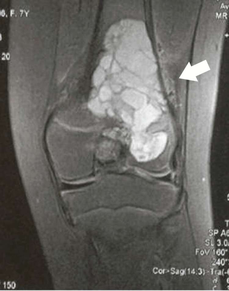
An aneurysmal bone cyst (ABC), when located juxta-physeal, may rarely penetrate the growth plate and extend into the epiphysis. The recurrence rate is considered higher when ABC is in contact with the active growth plate. Treatment methods usually focus on cyst healing and the rate of cyst recurrence. We present the method of treatment used [...]

Restoration of Limb Length Discrepancy and Alignment With the Ilizarov Device After Management of an Aneurysmal Bone Cyst Crossing the Distal Femoral Physis2026-07-08T09:49:21+03:00

Osteoid osteoma of the acetabulum: diagnosis and medical treatment

2026-07-08T09:46:22+03:00

We describe the medical management of an acetabular osteoid osteoma in an 11-year-old girl. There was a 2-year delay until proper diagnosis of the patient's pathology was made. Accurate localization of the nidus in the subchondral bone, involving the cartilage, is difficult on the basis of plain radiographs. All imaging methods, including computed tomography, MRI, [...]

Osteoid osteoma of the acetabulum: diagnosis and medical treatment2026-07-08T09:46:22+03:00

Diagnostic approach and arthroscopic treatment of septic arthritis of the knee, in an infant

2026-07-08T09:45:51+03:00

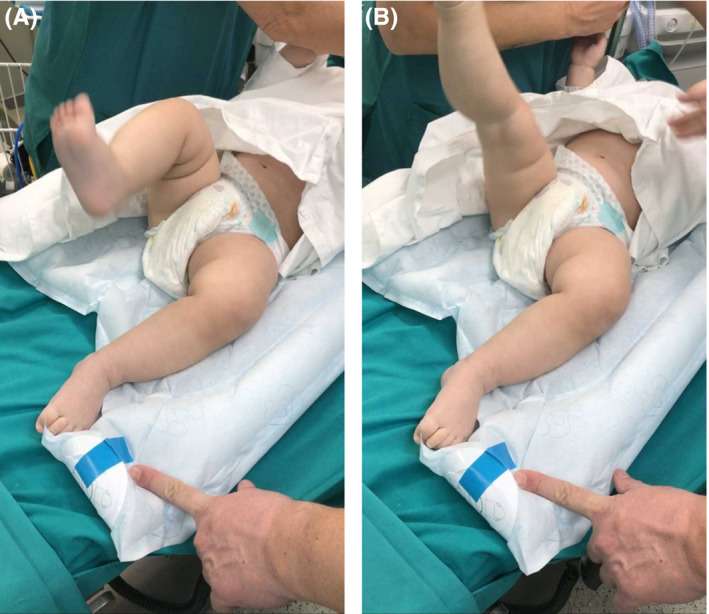
A 9-month-old baby presented with sudden inability to stand and unable to move his leg. Clinical examination showed edema and knee effusion. Blood tests and MRI confirmed septic knee arthritis without bone involvement. He was treated with arthroscopic lavage. He had a complete recovery and normal growth.   FIGURE 1 A, B Continuous kicking [...]

Diagnostic approach and arthroscopic treatment of septic arthritis of the knee, in an infant2026-07-08T09:45:51+03:00

Osteoid Osteoma in Children Younger than 3 Years of Age

2026-07-08T09:44:03+03:00

We present a case series of four children, younger than 3 years old, with osteoid osteoma of the lower limb. Pain and limping were the main symptoms. With careful clinical examination, we could localize the affected area. Radiological evaluation revealed cortical thickening in 3 children. On MRI examination, we found extensive edema, with normal bony [...]

Osteoid Osteoma in Children Younger than 3 Years of Age2026-07-08T09:44:03+03:00

Spontaneous union of bilateral congenital pseudoarthrosis of the clavicle, in a baby

2026-07-08T09:41:30+03:00

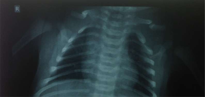
Congenital bilateral pseudoarthrosis is an extremely rare condition. We report a neonate with bilateral congenital clavicle pseudoarthrosis. The neonate had a palpable gap bilaterally. Radiological examination confirmed the diagnosis. The baby had a complete spontaneous healing in a year. We review the recent literature.   Fig. 1 Bilateral pseudarthrosis of the clavicle. Note the [...]

Spontaneous union of bilateral congenital pseudoarthrosis of the clavicle, in a baby2026-07-08T09:41:30+03:00

Neonatal Transphyseal Separation of the Distal Humeral Epiphysis: Early Diagnosis and Immediate Treatment with Radiographic Evaluation

2026-07-08T13:28:43+03:00

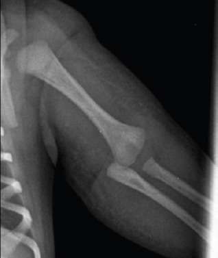
Introduction: Epiphyseal separation of the distal humeral epiphysis is an extremely rare injury. Both clinical and radiological examinations are unclear and delayed diagnosis is reported. The absence of the ossification center of the capitellum complicates the radiological diagnosis and often it is reported as elbow dislocation. Case report: We are reporting a case of a [...]

Neonatal Transphyseal Separation of the Distal Humeral Epiphysis: Early Diagnosis and Immediate Treatment with Radiographic Evaluation2026-07-08T13:28:43+03:00
Go to Top