orthopedikos

About admin

This author has not yet filled in any details.
So far admin has created 51 blog entries.

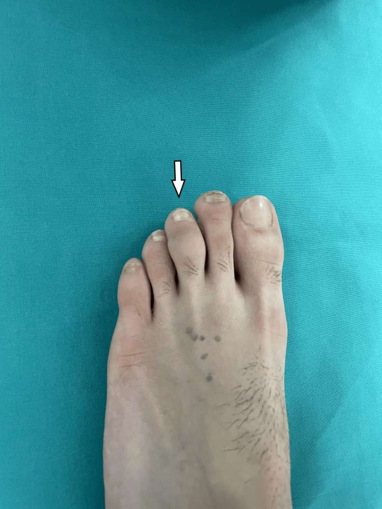
Sclerosing Intracancellous Osteoid Osteoma Presenting as a Painless Swelling of the Middle Phalanx in the Toe of a 15-Year-Old Footballer

2026-07-08T13:28:58+03:00

Osteoid osteoma (OO) most commonly affects the cortices of long bones and rarely involves the toes, where it typically presents with swelling and pain. Radiological evaluation can often pose a diagnostic challenge. We report the case of a boy with a painless swelling of the middle phalanx of the third toe. Radiographic assessment using plain [...]
Sclerosing Intracancellous Osteoid Osteoma Presenting as a Painless Swelling of the Middle Phalanx in the Toe of a 15-Year-Old Footballer2026-07-08T13:28:58+03:00

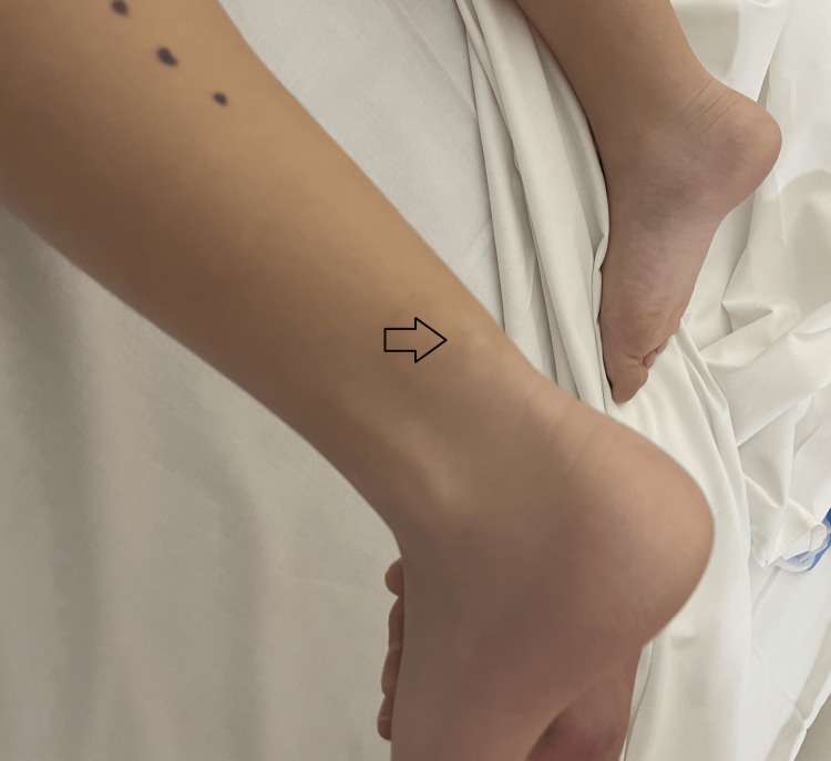
A Case Report of Deformity Correction of a Limb with Congenital Distal Tibiofibular Diastasis

2026-07-08T13:24:39+03:00

Introduction: Congenital distal tibiofibular diastasis is an extremely rare disorder. The main feature at presentation is a clubfoot deformity with leg length discrepancy (LLD) due to a short tibia. Limb lengthening and reconstruction techniques today can correct the deformity, ending with a plantigrade and functional limb. Case report: We report a patient with congenital tibiofibular [...]

A Case Report of Deformity Correction of a Limb with Congenital Distal Tibiofibular Diastasis2026-07-08T13:24:39+03:00

Perivascular Epithelioid Cell Tumor (PEComa) Affecting the Knee in a Seven-Year-Old Girl: A Case Report

2026-07-08T13:22:05+03:00

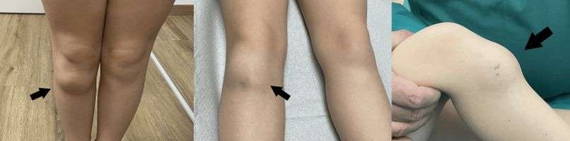
Perivascular epithelioid cell tumors (PEComas) are rare mesenchymal tumors composed of cells with epithelioid and spindle morphology, which grow around blood vessels. PEComas of soft tissue are usually reported in adults, with a rare incidence in the pediatric population, especially in young children and in the soft tissue of the limb. We report a case [...]

Perivascular Epithelioid Cell Tumor (PEComa) Affecting the Knee in a Seven-Year-Old Girl: A Case Report2026-07-08T13:22:05+03:00

Salter-Harris type II fracture of the distal femur in a newborn: Acute anatomic imaging alterations after labor dystocia

2026-07-08T13:18:04+03:00

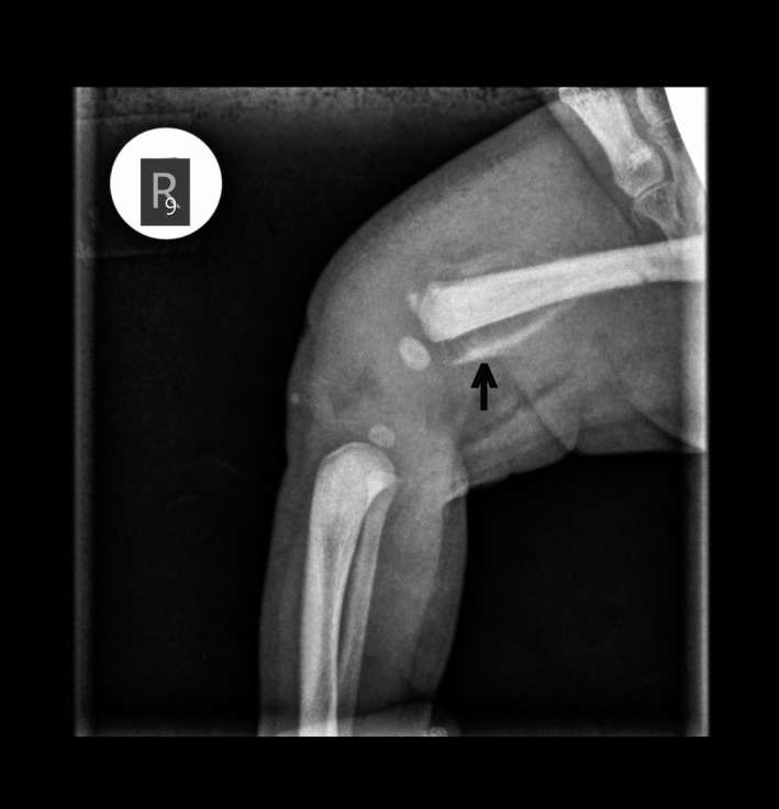
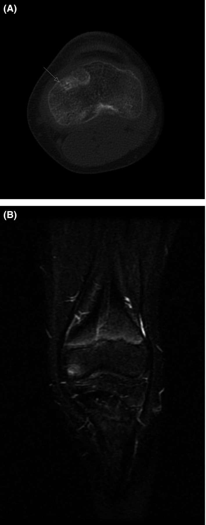
A rare clinical presence of Salter-Harris type II fracture of the distal femur in a newborn. The crucial role of imaging in depicting urgent anatomical alterations.   Figure 1 X‐ray of the right femur. The black arrow indicates the fracture and the detached osseous part Figure 2 MRI of the right femur [...]

Salter-Harris type II fracture of the distal femur in a newborn: Acute anatomic imaging alterations after labor dystocia2026-07-08T13:18:04+03:00

Schwannoma of the lumbar spine, presenting with pain of the knee, like an osteoid osteoma, in a 10-year-old girl

2026-07-08T13:14:40+03:00

Localized pain in the absence of local lesion may represent referred pain from the spine, in a child, arising from a benign spinal schwannoma. It can be diagnosed by MRI. Surgical excision of the tumor relieves the symptoms.   Figure 1 A‐B: Initial MRI and CT scan with an area of lucency with edema [...]

Schwannoma of the lumbar spine, presenting with pain of the knee, like an osteoid osteoma, in a 10-year-old girl2026-07-08T13:14:40+03:00

Solitary osteochondroma of the capitate, in a child

2026-07-08T13:11:53+03:00

Osteochondromas, are the most common tumors of the long bones in children. Osteochondromas are extremely rare in the carpal bones. They are diagnosed in adult life, in almost all cases in the literature. We report a 7 year old boy, who presented with a hard mass on the dorsum of his hand, with decreased wrist [...]

Solitary osteochondroma of the capitate, in a child2026-07-08T13:11:53+03:00

Bilateral femoral fractures during birth, in a neonate with malformation complex with stiffness

2026-07-08T13:09:36+03:00

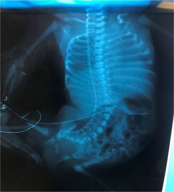
Fetuses with prenatal diagnosis of reduced mobility and malformation are at increased risk to sustain neonatal femoral fractures. Labor difficulties even after an elective cesarean section may trigger the fractures of the long bones. Figure 2 X‐ray showing bilateral femoral fractures. Casts were applied for fixed equinovarus feet Figure 3 X‐ray showing [...]

Bilateral femoral fractures during birth, in a neonate with malformation complex with stiffness2026-07-08T13:09:36+03:00

Figure 6. CT image on the axial plane showed a thin calcified lesion, located above the Achilles tendon, extending toward the sural nerve and vessel.

2026-07-08T13:07:22+03:00

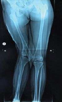
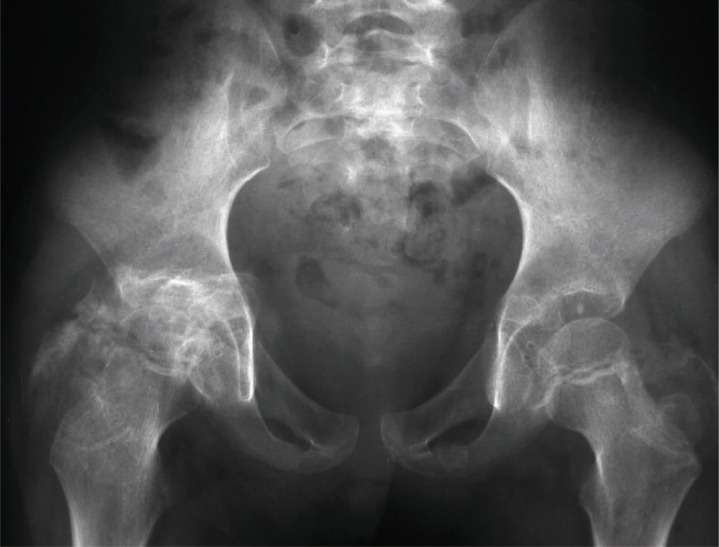
Introduction: Residual orthopedic deformities after meningococcal infection in infancy, including severe angular deformities and leg length discrepancy (LLD), affect patient quality of life. Case presentation: We report the case of an adolescent female with severe varus deformity and LLD of her right femur. We applied a preconstructed Ilizarov frame and initially corrected the varus deviation [...]

Figure 6. CT image on the axial plane showed a thin calcified lesion, located above the Achilles tendon, extending toward the sural nerve and vessel.2026-07-08T13:07:22+03:00

Fibro-Osseous Pseudotumor Presenting as Calcified Subcutaneous Mass Over the Achilles Tendon in a Five-Year-Old Girl

2026-07-08T13:03:14+03:00

A calcified mass located in the subcutaneous area above the Achilles tendon of a child poses difficulty for a correct diagnosis. Differential diagnosis includes tumoral calcinosis, calcified fibroma, calcified tendinitis, and myositis ossificans. A fibro-osseous pseudotumor is a rare benign lesion that usually affects the digits. Histologically, it consists of fibrous tissue with areas of [...]

Fibro-Osseous Pseudotumor Presenting as Calcified Subcutaneous Mass Over the Achilles Tendon in a Five-Year-Old Girl2026-07-08T13:03:14+03:00

Two cases of myositis ossificans in children, after prolonged immobilization

2026-07-08T13:01:07+03:00

Myossitis ossificans (MO) is a benign disorder characterized by heterotopic bone formation in skeletal muscle. It is divided in three types, fibrodysplasia ossificans progressive (FOP), myositis ossificans circumscripta or traumatica (MOT) and myositis ossificans without a history of trauma (non traumatic or pseudomalignant MO). Myositis ossificans is extremely rare in children younger than 10 years. [...]

Two cases of myositis ossificans in children, after prolonged immobilization2026-07-08T13:01:07+03:00
Go to Top