
ΕΠΙΣΤΗΜΟΝΙΚΟ ΕΡΓΟ
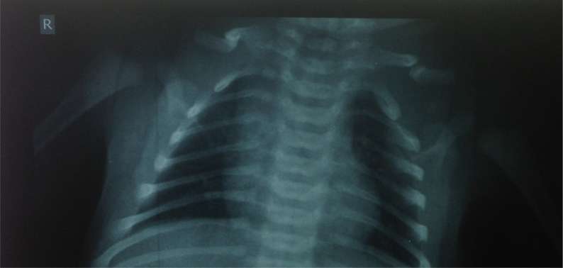
Spontaneous union of bilateral congenital pseudoarthrosis of the clavicle, in a baby
Congenital bilateral pseudoarthrosis is an extremely rare condition. We report[...]
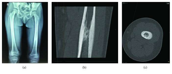
Osteoid Osteoma in Children Younger than 3 Years of Age
We present a case series of four children, younger than[...]
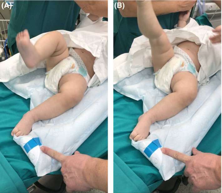
Diagnostic approach and arthroscopic treatment of septic arthritis of the knee, in an infant
A 9-month-old baby presented with sudden inability to stand and[...]

Osteoid osteoma of the acetabulum: diagnosis and medical treatment
We describe the medical management of an acetabular osteoid osteoma[...]
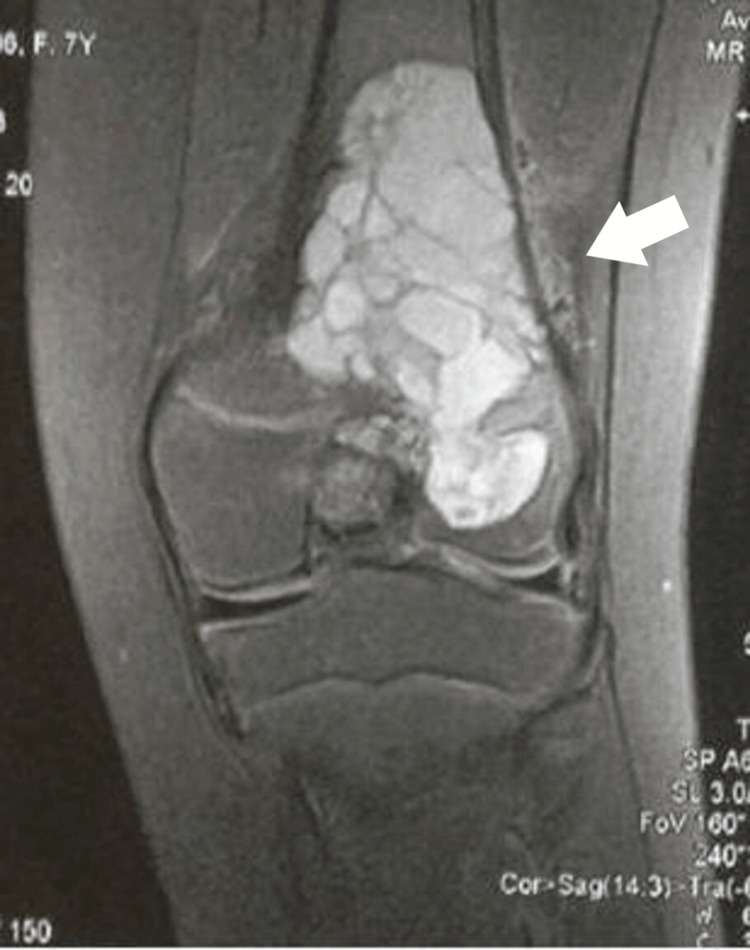
Restoration of Limb Length Discrepancy and Alignment With the Ilizarov Device After Management of an Aneurysmal Bone Cyst Crossing the Distal Femoral Physis
An aneurysmal bone cyst (ABC), when located juxta-physeal, may rarely[...]
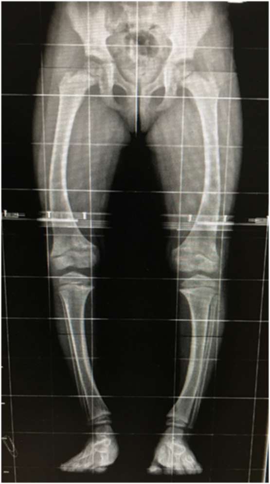
X-linked hypophosphatemia due to a de novo novel splice-site variant in a 7-year-old girl with scaphocephaly, Chiari syndrome type I and syringomyelia
X-linked hypophosphatemia (XLH) is a rare X-linked dominant inherited disorder[...]
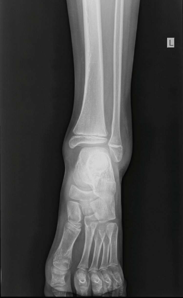
Displaced Distal Fibular Epiphysis in a Child: A Clinical and Radiological Evaluation of a Rare Injury Hidden From the Initial Radiological Examination
Fractures of the lateral malleolus are common in children. Isolated[...]
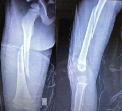
Impressive Remodeling of the Fractured Femur with Transposition of the Fractured Parts, Treated Conservatively with skin Traction, in a 3-Year-Old Boy
Introduction: Conservative management of femoral fractures in children in the[...]
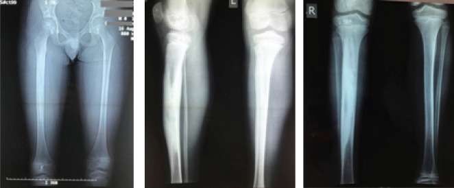
Melorheostosis in an Adolescent with Limb Length Discrepancy and Management with Epiphysiodesis with Eight Plates
Introduction: Melorheostosis is a rare bone dysplasia. It is mainly[...]
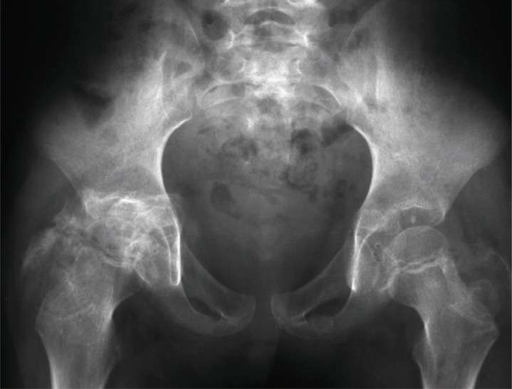
Two cases of myositis ossificans in children, after prolonged immobilization
Myossitis ossificans (MO) is a benign disorder characterized by heterotopic[...]
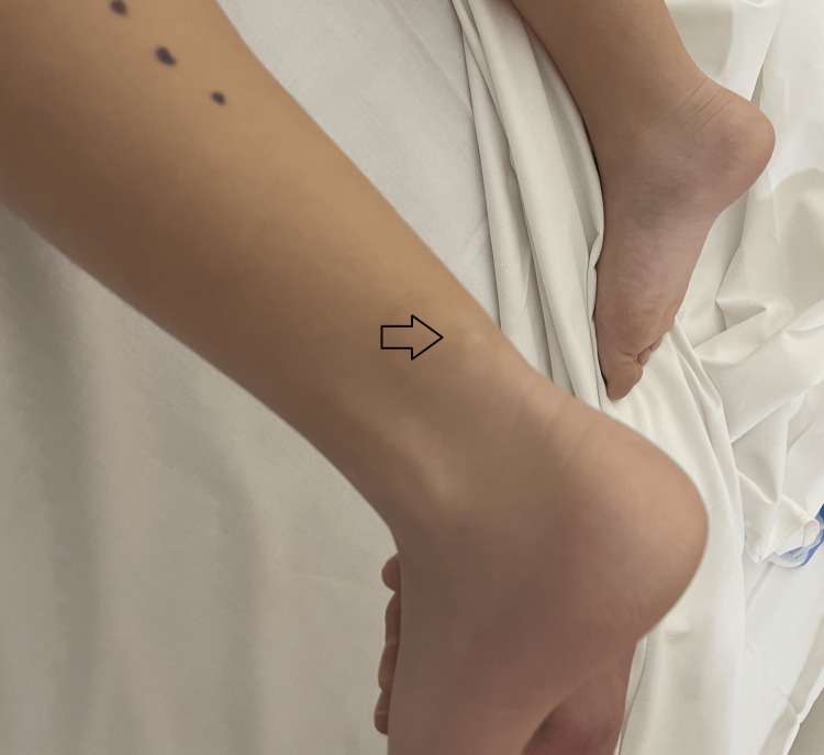
Fibro-Osseous Pseudotumor Presenting as Calcified Subcutaneous Mass Over the Achilles Tendon in a Five-Year-Old Girl
A calcified mass located in the subcutaneous area above the[...]
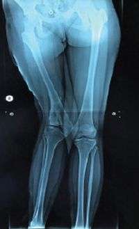
Figure 6. CT image on the axial plane showed a thin calcified lesion, located above the Achilles tendon, extending toward the sural nerve and vessel.
Introduction: Residual orthopedic deformities after meningococcal infection in infancy, including[...]